Admin_Adham
Knowledge of breast, nipple, and areolar (BNA) diseases is important for dermatologic care of all patients — and in those who are gender diverse, gender-affirming practices, such as chest binding and hormone-induced tissue changes, introduce additional diagnostic considerations.
“When evaluating a gender-diverse patient, the standard differentials for breast skin disease still apply, but there are unique dermatologic considerations and additional diagnostic pathways you should keep in mind,” Emily R. Nadelmann, MD, said at a conference on dermatologic care for sexual and gender minorities held at George Washington University (GWU). “These pathways add to the differentials — they do not replace them,” added Nadelmann, assistant professor, Department of Dermatology, GWU School of Medicine and Health Sciences, Washington, DC. Chest binding, for instance, can cause irritant or allergic contact dermatitis, koebnerization of psoriasis and other conditions, and infectious conditions such as candidiasis, scarring, and hyperpigmentation.
Yet, Nadelmann said, not all skin changes on the chest can be solely attributed to binding — a practice in which individuals compress the chest tissue for gender congruence. An eczematous dermatitis of the nipple that is unilateral and refractory to 4 weeks of treatment with topical corticosteroids, for instance, should prompt a biopsy for Paget disease. “It’s Paget disease until proven otherwise,” she said, referring to a hypothetical case of a 32-year-old transmasculine patient who presents with a unilateral scaly nipple erosion and attributes it to use of a binder.
With prolonged use, testosterone therapy can cause progressive lobular atrophy of the breast, though glandular tissue does persist, according to Nadelmann. Histopathologic studies have also found an association between testosterone therapy and the risk for Toker cell hyperplasia — a benign proliferation of clear cells in the nipple epidermis that can mimic Paget disease of the breast on pathology.
Despite the atrophic changes induced by testosterone, atypia and ductal carcinoma in situ have been identified in testosterone-exposed specimens from masculinizing mastectomy, Nadelmann said, underscoring the importance of routine pathologic evaluation of all surgical specimens.
Transmasculine top surgery leaves variable amounts of residual breast tissue, she noted, and the most common technique — double-incision mastectomy with free nipple grafting — transects the intercostal nerves, often resulting in diminished or absent nipple sensation.
Feminizing hormone therapy (estradiol and/or antiandrogens) promotes the formation of ducts, lobules, and acini, all of which are histologically similar to the cisgender female breast. “This is not gynecomastia....It is real breast tissue,” Nadelmann said.
Estrogen-driven decreases in sebum production can exacerbate xerosis and eczema, while the development of new breast tissue and inframammary folds can predispose to intertrigo and candidal dermatitis. The nipple and areola may also be susceptible to friction as new breast tissue develops, she said.
Breast cancer screening in gender-diverse patients should be individualized based on tissue present and on hormonal exposure history — not on gender identity alone, Nadelmann emphasized. “Dermatologists may be the only ones to detect suspicious chest skin changes [in gender-diverse patients] and to encourage them to pursue appropriate screening,” she said.
In situ and invasive breast cancers have been reported in transfeminine patients and may present at younger ages than in the general population, Nadelmann noted. According to current guidelines from the American College of Radiology, average-risk transfeminine patients who have received 5 years or more of gender-affirming hormone therapy should begin screening mammograms at age 40.
Transmasculine patients who have not had a gender-affirming mastectomy (GAM) should follow standard cisgender female breast cancer screening guidelines, and those who have had GAM should follow an individualized screening schedule, she said.
An Inclusive Chest Exam, More on Binding
An inclusive exam of the breast/chest should follow a conversation about pronouns and body terminology, Nadelmann said, and should involve stepwise exposure of the chest (examine, cover, and move on), sharing clinical reasons for the exam, and securing patient permission before binder removal.
Use “chest skin exam” unless the patient uses the term “breast exam,” she advised, and document the anatomy present and not assumed gender. For example, use “breast tissue present bilaterally, patient on testosterone therapy,” for instance, and do not use “female chest exam.”
“Always ask about binding, in a sensitive way, at every visit,” Nadelmann said. While many binding-related symptoms emerge within the first year, skin complications specifically tend to develop later, often after the patient has already begun experiencing pain.
In a 2017 survey study of 1800 individuals who had ever used chest binding, compression methods associated with the most symptoms attributed to binding were commercial binders, elastic (Ace) bandages, and duct tape or plastic wrap. To help patients manage risk, “we should counsel patients to use breathable materials and to avoid tape and Ace bandages,” Nadelmann said. Taking breaks and sizing up if one is in between sizes of commercial binders can also help reduce risk.
During a discussion session at the meeting, Steve Daveluy, MD, associate professor and dermatology program director at Wayne State University, Detroit, Michigan, and another speaker, said that chest binding takes “a little trial and error.” Sometimes, he said, “If patients can’t find a binder that’s good for the skin but tight enough, they can layer, with a wicking layer underneath a tighter binder…that sometime can help pull moisture away from the skin.”
Nadelmann advised being sensitive to cost differences of the various binding methods and patients’ varying budgets.
Hypertrophic Scarring After Top Surgery
In a 2024 review, hypertrophic postmastectomy scarring was found to occur in 17%-37% of cases of people who had undergone double-incision GAM. In addition, non-White race and perioperative androgen use were found in a recently published study to be significant risk factors for hypertrophic scarring following GAM.
Multiple clinical studies, including a review of patients undergoing GAM between 2017 and 2020 and a systematic review, have shown, however, that discontinuing testosterone perioperatively does not reduce scarring rates (or hematomas) compared with continuing it, Nadelmann said in an interview after the meeting.
“The effect of testosterone on scarring and hematoma risk likely reflects cumulative, long-term tissue-level changes rather than an acute pharmacologic effect that can be reversed by brief perioperative cessation,” she explained.
Preclinical data, she said, show that androgens induce pathologic scarring through dysfunctional extracellular matrix deposition (increased fibrosis, altered collagen ratios, and upregulated keratinization pathways), changes that would not reverse with a few weeks off hormones. Moreover, she added, discontinuation can “negatively impact mental health.”
Patients should instead be counseled about their individual risk profile for impaired wound health and hypertrophic scarring postmastectomy, with emphasis on optimized postoperative wound care and the use of “early scar prevention strategies,” such as silicone sheeting and compression, she told Medscape Medical News.
Intralesional triamcinolone and 5-fluorouracil can be used to treat hypertrophic scarring — just as they are used with other scarring and keloids — and laser treatment can be used for erythematous/early scars, Nadelmann said at the meeting. Silicone scar gel is another prophylactic option.
Complications From Unregulated Silicone Injections
Among populations of transfeminine patients, up to 50% of those seeking chest feminization have reported receiving unregulated silicone injections. Published dermatologic complications include silicone migration, inflammation, post-inflammatory hyperpigmentation, and infection.
When patients with a history of nonmedical silicone injections and cellulitis fail antibiotic therapy, consider atypical mycobacteria — and Mycobacterium abscessus, not just methicillin-resistant Staphylococcus aureus, Nadelmann advised.
Silicone vacuoles can also induce permanent blood diffusion inside circulating monocytes, she noted. And unregulated injections have been reported to lead to what’s called the autoimmune/autoinflammatory syndrome induced by adjuvants, in which local granulomas can evolve to Sjögren disease, sarcoidosis, and vasculitis. MRI is the preferred imaging modality for investigation, she said.
Complications from unregulated silicone injections are “important for derms to have on their differential, to ask about,” Nadelman said in the interview. Individuals “may not be open to disclosing it…but it’s a key part of their history for you to be able to help them.”
‘Do Not Miss’ BNA Differentials
Paget disease is one of what Nadelmann calls the “must not miss” triad of breast diseases with skin presentations that dermatologists must not overlook in any patient, including gender-diverse patients.
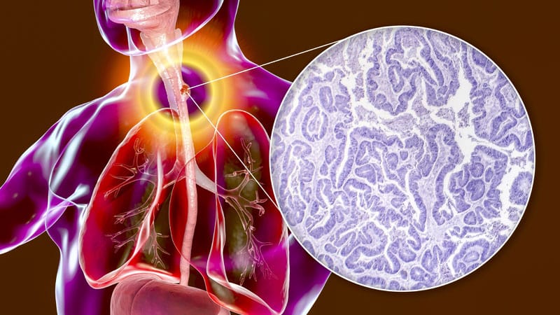
Paget disease usually presents unilaterally with variable involvement of the areola and a bloody/serosanguinous discharge and does not improve with topical corticosteroids. “Eight-five to ninety percent will have underlying cancer, and you’ll see pagetoid cells in the epidermis,” she said at the meeting.
Nipple eczema, on the other hand, is usually bilateral, involves the areola, and improves with 1-4 weeks of treatment with topical steroids. Discharge is rare, and histology will show spongiotic dermatitis.
“If the biopsy is benign but your suspicion is high, you should definitely rebiopsy and take deeper sections and check for CK7/CK20 immunostains,” she said.
Patients with inflammatory breast cancer (IBC) — another disease in Nadelmann’s don’t-miss triad — often present with rapid erythema and peau d’orange skin on a third or more of the breast. Some have what is thought to be mastitis that is not improving with treatment, she said. A punch biopsy does not rule out IBC, she noted.
Dermatologists should also be aware that radiation-induced angiosarcoma (RIAS) in patients with a history of radiation treatment for breast cancer typically presents 7-10 years after breast radiation therapy as violet lesions in the prior radiation therapy field. MYC gene amplification distinguishes RIAS from benign postradiation lesions.
In general, Nadelmann told Medscape Medical News after the meeting, the diversity of breast conditions with dermatologic presentations speaks to the importance of sensitive breast exams being part of total body skin examinations. “We lose diagnostic opportunities every time the chest is skipped — and it’s skipped more often than we think,” she said.
Granulomatous Mastitis: What Dermatologists Should Know
Somewhat underappreciated in dermatology — and important for gender-diverse patients, not only cisgender individuals — are granulomatous mastitis (GM) and “GMENA syndrome,” newly named to describe co-occurring GM, erythema nodosum, and arthritis, Nadelmann said at the meeting.
Patients with GM, a chronic inflammatory breast disease of unknown etiology and the “number one benign mimic of IBC,” are sometimes misdiagnosed as they shuffle between breast surgeons, infectious disease specialists, and dermatology, Nadelmann said in the interview.
Patients with GM present with painful breast masses, draining sinuses, and sometimes erythema nodosum, Nadelmann said. She saw many cases during her dermatology training and continues to see cases in the clinic — experience that has led her to believe that dermatology-led multidisciplinary care is beneficial.
GMENA syndrome is described in the literature as a rare entity associated with autoimmune rheumatic diseases, but Nadelmann said in the interview she believes it’s underdiagnosed and underreported, with patients sometimes presenting with erythema nodosum and having GM incidentally noted.
Nadelmann, who is also an assistant professor of dermatology at the University of Maryland School of Medicine in Baltimore, reported having no relevant disclosures.