Admin_Adham
It wasn't long after Iltefat Hamzavi, MD, began using ultrasound in his care of patients with hidradenitis suppurativa (HS) that he came to believe it was indispensable.

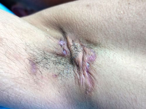
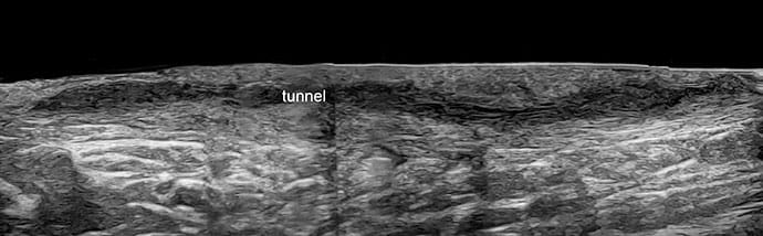
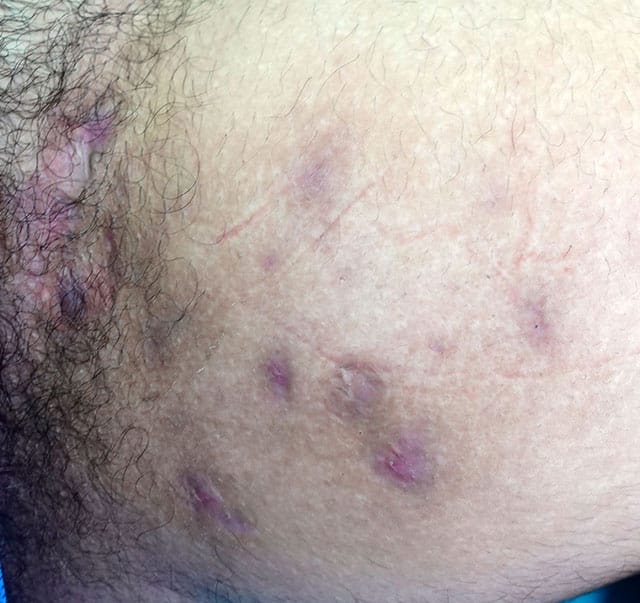
With the technology, he can detect and characterize subclinical lesions — fluid collections; fistulous tracts, also known as tunnels; and pseudocysts — that cannot be diagnosed by clinical examination alone. He can assess the extent of tunnels and map out lesional tissue before surgery. He can better understand the severity of disease.
"I don't think you can really [manage] HS very well without ultrasound anymore," Hamzavi, a senior staff physician at Henry Ford Hospital, Detroit, told Medscape Medical News.
Ultrasound has not yet made its way into clinical practice guidelines for the management of HS — at least, those used by clinicians in the United States. And while some of the HS clinics in the United States are now using ultrasound in the diagnosis and management of HS, it is "not yet the standard of care," as it is across parts of Europe and Latin America, said Hamzavi, who focuses on HS and pigmentary disorders.
But the literature is now replete with reviews of the use of ultrasound for HS, and multiple published studies have demonstrated the discordance between clinically and sonographically determined severity due to limitations of the clinical exam and its frequent underestimation of disease severity. Such studies have also documented high rates of subsequent changes in management and treatment.
And an international consensus statement published last year on the use of ultrasound in HS reported a high level of consensus among 24 experts (over half of whom were from dermatology departments) on using the tool to support diagnosis, staging, monitoring, presurgical planning, and research.
The experts — from 14 countries and four continents — universally agreed through a three-round Delphi study, for instance, that "ultrasound is needed in HS because the activity and severity of the disease are usually clinically underestimated," and that "ultrasound is required in all HS patients at all clinical stages."
Changes in Management
HS is "tremendously challenging to measure," said Hamzavi, who practices in the Detroit area and was one of three experts from North America who participated in the international Delphi study.
"There are a lot of biases introduced based upon pain, drainage, [and other parts] of the patient experience," he said. "Ultrasound can help reduce that bias, and it can sometimes help validate what the patient tells us."
It can also "reduce inter-observer variability," he said. A 2022 study of 22 patients that Hamzavi co-authored showed that the addition of ultrasound evaluation improved inter-rater agreement of various HS outcome measures.
Ultrasound has been helpful for presurgical planning, Hamzavi emphasized, and in his patients, it has "helped reduce the healing time because we're able to be more specific about which areas we excise. … We can precisely map out where the tunnels are." A study recently submitted for publication documents this reduced duration of healing, he said.
(The international consensus statement supports the role of ultrasound in HS surgery, with 100% agreement that peri-operative ultrasound can help improve recurrence rates in surgery, and that recurrences of already excised lesions should be evaluated with ultrasound.)

While Hamzavi has been using and studying ultrasound in HS for over 10 years, Hadar Lev-Tov, MD, who directs the Miami HS Center at the University of Miami School of Medicine, Miami, Florida, learned and incorporated the technology just several years ago, when portable, cordless, high-resolution ultrasound units became more available.
"You can now get a probe for about $5000, which is not cost-prohibitive when you consider other technology that sits in the dermatologist's office," Lev-Tov said.
His staff prepares the patient for an ultrasound assessment and connects the screen and probe so that, from the time Lev-Tov enters the exam room, ultrasonography often adds "only a minute or two to the assessment," particularly when the goal is to localize a suspected tunnel.
Aesthetics may be the area where the use of ultrasound is "picking up the most steam" in dermatology practices in the U.S., Lev-Tov told Medscape, referring to ultrasound guidance for filler injections, for instance. But in the clinical arena, "clearly HS is the lowest-hanging fruit in terms of impact," he said. "It's an obvious need."
More often than not, the addition of ultrasound will "upstage the disease," he said, "but sometimes it doesn't. Sometimes I look and think, 'Oh, that's not what I thought.'"

The low concordance of clinical and ultrasonographic findings was documented in 2013 in an oft-cited paper authored by Chilean radiologist Ximena Wortsman, MD, and colleagues. Wortsman, who has dedicated her research to ultrasound in dermatology and has written several textbooks on the subject, reviewed the sonograms of 34 consecutive patients and organized their abnormalities into a newly devised sonographic scoring system (SOS-HS) — inspired by the clinically based Hurley staging system — with most relevant anatomical sonographic findings.
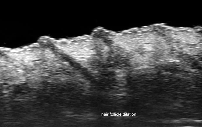
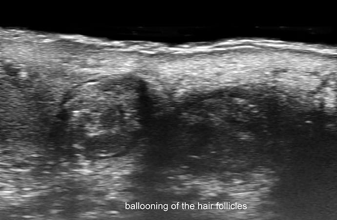
Clinically unrecognized fluid collections were observed in 76% of patients, fistulous tracts in 30%, dermal pseudocysts in 71%, and widening of the hair follicles in 100%. Patients were staged according to the new SOS-HS.
Notably, Wortsman, the medical director of the Institute for Diagnostic Imaging and Research of the Skin and Soft Tissues in Santiago, Chile, documented that the management of more than 80% of the cases had been modified after ultrasound examination, with different medications used or treatment switched from medical to surgical approaches, consistent with the new sonographic scores.
"If you use ultrasound, your management will change in more than 80% of the cases," she told Medscape Medical News. "Why? Because we changed the stage … we learned, for instance, that what was thought to be healthy skin between two lesions was not really healthy."
Other studies have similarly shown how ultrasound can modify the clinical staging and therapeutic management of HS by detecting subclinical disease. In a cross-sectional multicenter study in Spain involving 143 patients, for example, most cases were switched to a more severe Hurley stage classification after ultrasound, which subsequently modified the therapeutic approach. Of those with a stage I classification specifically, 45% were changed to a more severe stage.
An Italian ultrasound working group comparing clinical and ultrasound scores similarly reported in 2019 that 29% of patients were classified in a more severe stage after ultrasound assessment. And in a 2016 report by Wortsman and colleagues on ultrasound diagnosis and staging in pediatric HS, management was modified after ultrasound in 92% of cases.
Learning Curve and Evolving Technology
Learning how to use ultrasound for HS is fairly straightforward, Lev-Tov and Hamzavi both said. "My guess is that if you do a 1- to 3-hour class and then use it regularly, correlating the clinical exam with ultrasound results, within 2 to 3 months, you'll be very comfortable," Hamzavi said in the interview.
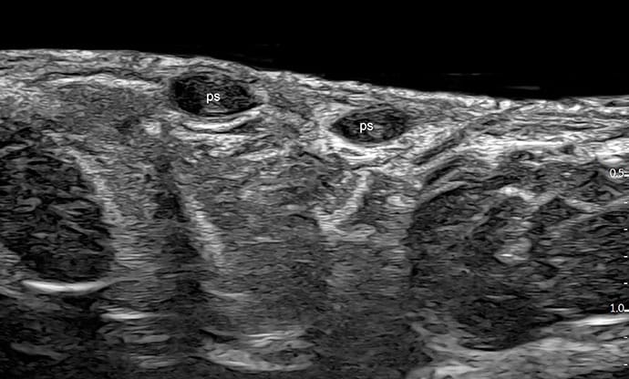
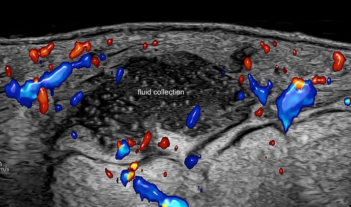
The international consensus statement solidifies the "new language" needed for describing and defining key lesions in HS, he said. Ultrasound fluid collections in HS, for example, are defined as hypoechoic or anechoic dermal and/or hypodermal sac-like structures that measure ≥ 1 cm. Pseudocysts are round or oval-shaped hypoechoic or anechoic dermal and/or hypodermal sac-like structures measuring < 1 cm.
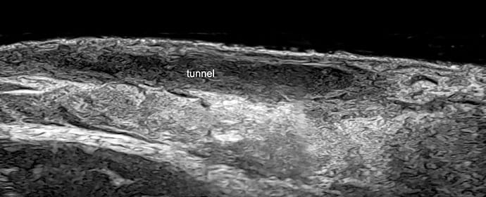
And ultrasound tunnels in HS are defined as hypoechoic or anechoic band-like structures parallel to the skin surface, which, like the other two lesion types, are connected to the dilated base of the hair follicles. Tunnels, the statement notes, can show hypoechoic or hyperechoic laminar patterns in their periphery suggestive of fibrosis.
Dermatologists have lagged behind other specialists in their exposure to ultrasound during training, "but they'll pick it up quickly," Hamzavi said. "It's like the early years of dermoscopy — we knew it would do something and we learned it, and now we do it really well and it helps so much."
Experts agree that to perform an ultrasound exam in HS, the dermatologist should use an ultrasound probe with a minimum upper frequency of 15 MHz. Such high-frequency ultrasound allows one to visualize the dermal and subcutaneous skin layers and distinguishes lesional from non-lesional areas. "With 15 MHz, you can discriminate structures measuring 0.1 mm or more," Wortsman said, noting that MRI discriminates structures measuring 4 mm or more.
Color Doppler is also valuable for HS, she said, because it allows one to see vascularity and to monitor the inflammatory activity of HS lesions.
Ultra-high-frequency (>50 MHz) ultrasound can better detect early signs of HS, most participants of the consensus statement agreed, and particularly at 70 MHz, it can better detect the dilation and ballooning of hair follicles and other dermal abnormalities. "New ultra-high-frequency devices provide a resolution of up to 30 microns, which is almost a histological picture," Wortsman said.
Ultra-high-frequency ultrasound may be part of Lev-Tov's future, he said, where it could be potentially useful for even more "subtle" disease like skin cancer. But for now, his portable 20 MHz probe with a color Doppler option serves his patients well, he said.
Wortsman recently updated her system for sonographic scoring of HS severity — called the modified SOS-HS (mSOS-HS) — and devised a new system for scoring disease activity (separate from severity) based on abnormalities detectable using 70 MHz.
Tunnels themselves can also be classified according to the degree of sonographically visualized fibrosis and edema and according to their layer location and complexity. Research has shown that tunnels with more fibrosis "will probably be bad responders to biologic therapy," Wortsman noted.
Scoring and classification systems are currently used in multiple countries in daily practice, but not in the US. "I don't use them personally … but I think we're evolving toward [using them]" as the use of ultrasound in HS becomes more widely adopted, Hamzavi said.
Dermatological ultrasound training is available from organizations such as the American Institute of Ultrasound in Medicine, and sessions on ultrasound imaging of HS have also been available at annual meetings of the North American HS Foundation and other organizations.
In addition, Wortsman — who is a voluntary associate professor in dermatology at the University of Miami, where she is collaborating with Lev-Tov and other faculty on research — teaches a course on basic concepts of dermatologic ultrasound through the American Academy of Dermatology Learning Center. (The course is offered under the umbrella of skin cancer.)
Ultrasound of the skin has gained interest not only in Europe, but in Latin America and Asia, she said. "In some countries, radiologists are doing the exams for HS. In other countries, dermatologists are. It depends on who takes the lead and invests the time," said Wortsman, who is also an adjunct professor in the departments of dermatology at the University of Chile and the Pontificia Universidad Catolica de Chile, Santiago.
"In many places in Spain, for example, dermatologists have a machine in their department, and they have mornings or afternoons when they're dedicated to ultrasound," she said.
Pain generated by an ultrasound is infrequent in patients with HS, she said, referring to the results of a 2023 multicenter cross-sectional study of 317 patients with HS.
'Opportunities to Learn More'
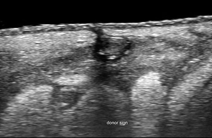
Ultrasound has, over the years, deepened understanding of the pathophysiology of HS by revealing the dilatation and then rupture of hair follicles, and the spread of fragments of hair tracts through deeper dermal or subcutaneous layers. It appears through ultrasound, Wortsman added, that different lesion types are generated based on differing patterns of fragmentation of keratin.
Lev-Tov sees firsthand how ultrasound "unveils opportunities to learn more." He recently led a review of studies of ultrasound-guided intralesional triamcinolone injections for HS lesions that produced "early data" suggesting there may be benefits in terms of response, healing, and pain. However, the review also made it clear that more research is needed to learn about optimal injection placement; injection location varied in the studies based on lesion type and provider preference.
"With eyes under the skin, we see that we now have choices, and questions. Is injecting in the middle of the tunnel OK, or should I give a certain dose every cm of the tunnel?" Lev-Tov said. "Or when injecting an abscess, should I fill it like a balloon or go to the base and inject the underlying dermis?"
Wortsman and other experts are pushing for ultrasound to be included in clinical trials of medications for HS. Such a recommendation was one of the 80-plus statements included in the international consensus paper.
And with time, data on the longer-term patient outcomes associated with routine incorporation of ultrasound in HS may emerge. "We have data that it changes management, but regarding overall outcomes for the long term — data that looks holistically — I don't know," Lev-Tov said. "But I have no doubt it improves patient outcomes."
"Right now," he said, "we need to popularize it, let doctors use it, and they'll see the value."
Lev-Tov and Hamzavi reported having no relevant financial disclosures. Wortsman reported receiving royalties from Springer Books, speaker fees from UCB and Novartis, and consultant fees from Galderma.