Admin_Adham
AUSTIN, Texas — The use of two-photon fluorescence microscopy is an innovative, emerging technology that is more accurate than frozen sections for Mohs surgery, according to research presented at the annual meeting of the American College of Mohs Surgery (ACMS) Annual Meeting 2026.
“We saw noninferiority in sensitivity, and we saw higher specificity than with frozen sectioning, leading us to conclude that two-photon fluorescence microscopy was more accurate than frozen sections for Mohs surgery,” Sherrif F. Ibrahim, MD, PhD, of Rochester Dermatologic Surgery in Victor, New York, told attendees.
“It is the first intraoperative slide-free histology study that shows noninferiority and possible superiority to standard frozen sections, and it had a potential direct impact on patient care,” he said, noting this work was done in collaboration with Michael Giacomelli, PhD, associate professor of biomedical engineering and optics at the University of Rochester, Rochester, New York.
The use of this technology could potentially transform the Mohs lab, Ibrahim said, with no need for sectioning, no cryostat, no embedding, no dedicated histology tech, and no block facing, with every margin evaluated accurately within an approximately 20-minute workflow “from the time we remove the tumor until the time that you get an answer.”
Two-photon fluorescence microscopy uses a near-infrared femtosecond laser to use two-photon excitation that has a highly specific, fine focal point that can be moved up and down, Ibrahim explained. This essentially allows for “optical sectioning” that can provide high-resolution histology in minutes without slides, obviating the need for frozen section preparation.
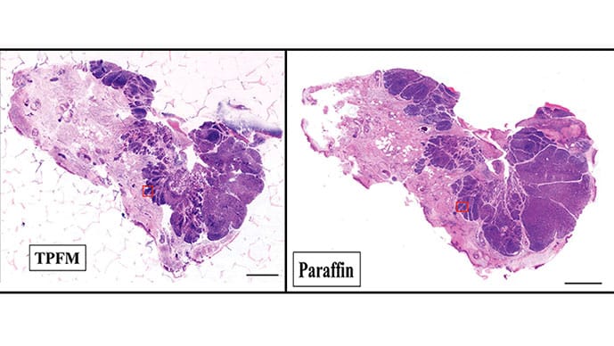
The tissue is removed from the patient and immediately undergoes a 3-minute nuclear stain to create a virtual hematoxylin and eosin (H&E) rendering of fresh, unprocessed tissue that provides images of the true surgical margin, not a processed section displaced from the cut edge, Ibrahim said. He explained how this microscopy is superior to confocal reflectance microscopy.
“Confocal is single-photon excitation using a much shorter wavelength, so there is a lot more scatter and, as a result, a shallower depth of penetration, so it’s not optimal for Mohs margin evaluation,” he said. The near-infrared excitation of two-photon fluorescence microscopy, meanwhile, results in less scatter, deeper penetration, and H&E-quality nuclear and stromal detail that confocal reflectance microscopy cannot provide. With two-photon fluorescence microscopy, there’s no physical sectioning of the tissue, no processing, and it’s nondestructive, so the tissue is preserved for permanent sections later, if desired.
Ibrahim then showed the point-of-care system, which has a similar footprint to a standard cryostat, and described the workflow used after they validated the technique.
After excision, staining, and mounting, microscopy processing took 2-3 minutes to evaluate margins. Then they gave the specimens to the histology technician for standard Mohs sectioning, “removing some portion of the skin and then generating what we see every day in most slides, which is essentially a pseudo margin,” Ibrahim said. Then they brought the specimen back to the two-photon system and imaged it again to get good co-registration with the frozen section.
To assess its accuracy for Mohs, the researchers compared the diagnosis of surgical margins on two-photon fluorescence microscopy with that from frozen section histology in 122 consenting patients with 169 Mohs margins from May 2024 to June 2025.
Ibrahim showed several comparisons of the two-photon fluorescence microscopy imaging vs frozen section imaging. “You couldn’t make any decision based on the two-photon fluorescence microscopy because that would change the standard of care,” he said, but they could compare them after the fact.
They found a false positive in 11 of the 122 first stages, “meaning we didn’t see tumor at the true margin,” Ibrahim reported. “Once we started sectioning deeper into the block, we did reveal tumor.”
That suggested that 25% of additional stages taken under frozen sectioning may be medically unnecessary, Ibrahim said. If that’s representative at a larger scale, it means up to 140,000 people may have unnecessary Mohs stages taken annually, he said.
Overall, two-photon fluorescence microscopy had higher accuracy than frozen sections, he reported. Among 169 specimens, two-photon fluorescence microscopy had 33 true positives and 132 true negatives compared with 32 and 123, respectively, for frozen sections. There were three false positives with two-photon fluorescence microscopy compared with 12 with frozen sections, as well as one false negative with two-photon fluorescence microscopy compared with two with frozen sections.
That translated to an accuracy of 97.6% with two-photon fluorescence microscopy, with 97.1% sensitivity and 97.8% specificity compared with 91.7% accuracy (P = .015) with frozen sections, which had 94.1% sensitivity (P = .0047) and 91.1% specificity (P = .017).
Nicole Negbenebor, MD, dermatologic surgeon and assistant professor of Mohs micrographic surgery and clinical oncology at the University of Iowa Carver College of Medicine in Iowa City, Iowa, told Medscape Medical News that she was impressed with how this technology could improve the accuracy of pathology, especially given the artifacts that can show up.
Mistaking a hair follicle for a basal cell, for example, could lead surgeons to take another layer, which adds costs, she said. “But if you could prove that it was a benign finding, you don’t need to take another layer in the Mohs stage, which is huge,” she said. It will be important to assess the upfront costs of the technology, but it seems that it could be cost-effective over time, she added.
Ibrahim reported having no disclosures. Negbenebor reported serving on an advisory board for UCB and serving as a clinical trial investigator for Johnson & Johnson and Sanofi.
Tara Haelle is a science/health journalist based in Dallas.