Admin_Adham
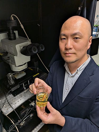
Late one night at Kyushu University in Fukuoka, Japan, Shigenori Inagaki, PhD, was stuck. For a year, he had been testing spherical polymers to make living brain tissue transparent, an idea his supervisor, Takeshi Imai, PhD, had essentially given up on a decade earlier. The best candidate, a synthetic polymer called Ficoll, looked promising on paper, but in practice the solution was too viscous, and it dampened neural activity. He had screened more than 100 compounds. Nothing worked well enough.
Then a thought arrived with the simplicity that only seems obvious afterward: proteins are polymers.
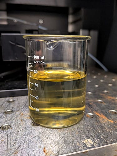
Inagaki grabbed a bottle of bovine serum albumin (BSA) from the shelf. Not just any BSA. This was the expensive, high-purity kind he wouldn’t normally use. He dissolved it in artificial cerebrospinal fluid (CSF), replaced the culture medium, and watched the cells immediately go transparent. He tested it three more times before he believed it.
Light scatters in tissue because the fluid between cells and the material inside them bend light differently. Match those optical properties and the tissue becomes transparent.
“The transparency itself was not surprising,” Inagaki said. “If you adjust the refractive index, tissue becomes transparent. That was already known. The most surprising thing was the osmolarity [concentration of particles]. It was ten times lower than Ficoll.”
That difference, invisible to the eye but lethal to cells, was the whole problem. And a protein already circulating in every mammal’s bloodstream had been the answer all along.
The Decade-Long ‘Impossible’ Problem
The study, published in Nature Methods and co-led by Inagaki and Imai, describes SeeDB-Live, the first tissue-clearing technique that works on living mammalian tissue without disrupting cellular function. It’s the third generation of Imai’s clearing platform, though calling it an update undersells what happened. The original SeeDB, published in 2013, used fructose to clear fixed, dead tissue for anatomical imaging. SeeDB2 followed in 2016, optimized for super-resolution microscopy. These tools took still photographs of the brain. But SeeDB-Live captures live videos.
Imai had been asked whether live tissue clearing was possible roughly a hundred times over those years, and he always said no. When asked whether there was a point during the decade when he stopped thinking about it, his answer was blunt: “I totally gave up.”
An earlier postdoc in his lab had tried and failed. Large laboratories in the US and Europe had tried and failed. Why had this problem proven itself to be so intractable? For one, tissue transparency isn’t the actual problem; making tissue transparent is straightforward optics. The problem was doing it without killing anything.
Small molecules like sugars can raise the refractive index of the extracellular fluid, but you need enormous concentrations to get there; the original SeeDB used fructose solutions nearly as thick as honey. This raises the osmotic pressure of the solution, pulling water out of a cell when the surrounding fluid is too concentrated. At the concentrations needed to clear tissue with sugars, that pull is strong enough to kill neurons.
Osmotic pressure scales with the number of dissolved particles, not size. What Inagaki recognized was that a very large molecule could raise the refractive index with far fewer particles, keeping osmotic pressure low enough for cells to survive. Albumin fit that description perfectly. It’s large, spherical, highly soluble, and has already been proven safe in the body. One albumin molecule does significant optical work but counts as just one particle for osmolarity.
But there was a catch. “Later on, we tested many kinds of BSA products from many vendors,” Imai said. “Only the high-purity BSA has extremely low osmolarity.” Crude BSA contains salt contaminants that raise ion concentrations and disrupt neuronal function. Had Inagaki reached for a cheaper bottle that night, the idea might have died on the bench. They later engineered a protocol that works with crude BSA, but the initial discovery required pure BSA, which is too expensive for extensive work.
Imai credits Inagaki’s engineering background as the reason it worked where others had failed. Where earlier attempts had relied on rough estimates, Inagaki precisely identified the refractive index sweet spot for living cells — between 1.36 and 1.37.
“Many laboratories tried over 10 years,” Imai said. “The difference in our success comes from Shigenori being very careful on the precise measurement of living cells.”
What You Can See
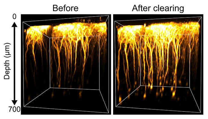
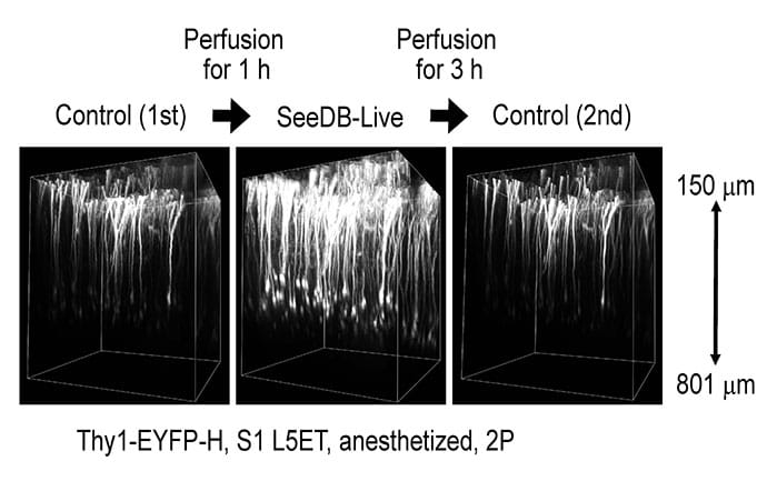
Mouse brain slices clear within an hour of immersion, confocal imaging depth roughly doubles, and deep neurons become about three times brighter under two-photon microscopy. Using patch-clamp recording, which measures the electrical activity of individual neurons through a tiny glass electrode attached to the cell membrane, the researchers confirmed that resting membrane potentials, action potential thresholds, and firing rates were essentially unchanged after clearing. Inagaki noted that some electrophysiologic parameters shifted slightly, but “the final output doesn’t change.”
The technique also works in living mice. Through a cranial window over the somatosensory or visual cortex, SeeDB-Live illuminated layer five cortical neurons at depths of 600-800 µm, with no detectable toxicity to neurons or behavior even after 120 days of repeated use. Once the albumin is washed out, the brain returns to its normal opacity within hours.
For Imai, the most exciting application may be voltage imaging. Most neuroscientists rely on calcium imaging to watch neural activity, largely because it’s easier, but calcium is an indirect readout, a downstream echo of the electrical signal. “Membrane potential is the true signal propagating in neural circuits,” Imai said. “It’s been really difficult to see.”
Combined with a high-speed camera, SeeDB-Live enabled widefield voltage imaging of dozens of dendrites simultaneously in a living mouse brain, capturing population-level electrical dynamics that were previously out of reach.
The technique also cleared spheroids and organoids, preserving calcium transients in intestinal organoids and normal growth in cortical organoids even when cleared for 4 hours daily. For researchers using patient-derived brain organoids to model neuropsychiatric disease or screen drug compounds, the ability to image deep functional activity in living tissue rather than fixing and killing the sample could fundamentally change what is measurable.
What It Can’t Do Yet
Delivery remains the largest limitation. In living animals, albumin diffuses only into the superficial cortex and can’t penetrate the blood-brain barrier if injected systemically. The CSF washes it away almost as fast as it diffuses in.
“We don’t know much about how the cerebrospinal fluid circulates within the brain,” Imai said. “If we knew the entire stream, we could make use of it. But we don’t fully understand it.” Inagaki is now working on CSF-based delivery strategies, introducing albumin through the ventricular system rather than relying on passive diffusion. Myelin-rich brain regions and subcortical structures like the hippocampus remain out of reach for now.
When asked whether topical application during open neurosurgery could enhance fluorescence-guided imaging of tumor margins, an obvious translational question given that albumin is already used clinically, Imai was cautiously open. “In theory, it should be possible, for example, to light up glioma,” he said. “But we are using very high concentrations of BSA. We need to develop a more efficient strategy.”
Annie Kathuria, PhD, an assistant professor of biomedical engineering at Johns Hopkins University who develops multi-region brain organoids (MRBOs) for neuropsychiatric and cancer research, said the technique could have immediate applications for tumor organoids. Her lab builds glioblastoma organoids and mixes them with healthy cerebral tissue to study how tumors invade surrounding brain — a model she said could benefit directly from live clearing.
“If we have one transparent organoid, along with one that isn’t, we can track how fast glioblastoma penetrates healthy tissue,” Kathuria said. Because glioblastoma organoids have less lipid than healthy brain tissue, she expects them to clear more readily, allowing researchers to watch drug efficacy and toxicity in real time, tracking which cell types are being killed and at which dose. “You can find the sweet spot,” she said.
But Kathuria cautioned that the technique faces a significant hurdle in human-derived brain organoids. Human brain tissue has far more lipid content than mouse, and the structure of human myelin is different than in mice. “If they tried using this technique in a human brain organoid, it might not work,” she said. She called the animal work “amazing and excellent,” but said the next step is demonstrating that SeeDB-Live performs in the human-derived models that drug screening ultimately depends on.
The Curious Side Project
Inagaki never expected to be here. Because he has an engineering background, he wanted to build microscopes and fluorescent probes. Every week he brought Imai new ideas during their lab meetings, but Imai rejected nearly all of them. Live tissue clearing was one of the few he approved of, and even then, he remained skeptical.
“I wasn’t so optimistic,” Imai admitted. “But Shinegori is very precise. That was the reason for his success.”
When asked whether he could have predicted 5 years ago that he would be clearing live brain tissue, Inagaki’s answer was simple: “I didn’t expect this.”
The field of tissue clearing has historically given neuroscience exquisitely detailed maps of dead brains. SeeDB-Live doesn’t replace those maps, but it offers something the field has never had: a way to watch the living brain at depths it couldn't reach before, using a protein that evolution put there long before anyone thought to look.
Kathuria reported having no relevant financial disclosures. Johns Hopkins Technology Ventures supported the commercialization of her MRBO technology. Disclosure information for study authors is available in the original study publication.