
 Admin_Adham
 Jump to Key Points
The radiology department is seeing cardiac tissue a whole lot clearer with the emerging use of photon-counting CT (PCCT) scanners. That means better diagnosis, better prevention, and broader eligibility for patients with cardiac disease who were previously unscannable, according to experts.
“We are coming into a new era in accuracy, and the implications are profound,” Arthur Shiyovich, MD, from Brigham and Women’s Hospital and Harvard Medical School in Boston, told Medscape Medical News. Most importantly, that accuracy will significantly reduce downstream testing for cardiac disease, he said.
In his recent editorial in JACC, Shiyovich described PCCT as “transforming cardiac CT from a primarily anatomic modality into a multidimensional imaging platform capable of integrating coronary assessment, cardiac structure and function, and myocardial tissue characterization.” For patients with implanted devices or claustrophobia, or an inability to stay still for longer scan times, or who are on a long waitlist, PCCT is a new option, “and may be further enhanced by emerging deep learning-based reconstruction and analysis techniques,” he added.

Conventional CT machines have improved with higher speed, lower radiation, and larger coverage, but detection has not improved because of electrical signal loss during two-step image conversion, whereby an x-ray must first be converted to light and then into an electrical signal. For patients with dense calcium, coronary stents, or other metal implants, the signal loss during conversion results in image distortion, causing “blooming” and blurring artifacts, which makes scans difficult to read. These patients are not considered good candidates for CT scanning and instead are sent to the cath lab for stress tests or scanned using other modalities (such as MRI) that involve lengthy wait times.
- PCCT improves coronary plaque/stenosis assessment; ↓ blooming, ↑ spatial resolution.
- Dense calcium, stents, metal implants now more interpretable; fewer “unscannable” patients.
- Studies show CAD-RADS downclassification ~34% and fewer downstream invasive CAG referrals.
- PCCT + FFR-CT may identify nonobstructive disease despite CAC >1000, reducing PCI.
- Limits: contrast still needed; ultrahigh-res scans may need comparable radiation; high cost/infrastructure.
PCCT scanners count photons and convert them directly to an electrical signal, making it possible to identify tissue. Each photon is then sorted into a so-called “bin,” making it possible to construct images that include or exclude stents, metal, plaque, calcium, muscle and other tissue, or implants. The technology makes heart vessel boundaries appear clear, identifiable, and measurable and high-risk fatty plaque visible behind calcification, research showed.
What’s more, PCCT is capable of capturing a very thin slice of tissue, 0.2 mm, compared with 0.6 mm for conventional CT, so objects very close together can now be viewed separately and measured more accurately.
“Everyone in the field is really excited about this technology,” Todd C. Villines, MD, from the University of Virginia in Charlottesville and executive editor of JACC: Advances, told Medscape Medical News.
“Our biggest challenge is artefacts and blooming…. This is really going to reduce inappropriate trips to the cath lab as we become more accurate and confident.”

Villines said the scope of advancement that PCCT technology offers continually surprises specialists. During SCCT2026, a “Stump the Masters, Stump the Professor” case involved a patient with cardiac disease who was sent to the cath lab whose tests came back normal. “All of us focused on the main artery,” Villines explained. However, when they looked at the patient’s PCCT scan, “we saw one of these tiny little septal branches was occluded — those are usually too small to interpret.”
“The PCCT was more accurate than CAG [coronary angiography] and better than invasive angiography by a lot; this really highlighted the strength of this technology,” he said.
“We now know when we call it a stenosis, you actually have a stenosis — particularly for those patients with stents.” Ultimately, Villines said that PCCT will lead to fewer complications and inappropriate procedures and better patient care.
Diagnosis Downgraded With PCCT
New data showed that PCCT is more accurate in quantifying plaques, reducing overdiagnosis. Conventional scanners overestimate plaques because of “blooming,” as reported in a 2025 article in Radiology. In 48 patients scanned, lower plaque volume was seen with ultrahigh-resolution PCCT than in energy-integrating detector CT (724 mm3 vs 10,853 mm3). Better reproducibility was also reported using PCCT.
The same researchers reported decreased overestimation of luminal narrowing with dense calcification and lower plaque-measured stenosis when scanning with PCCT vs conventional CT, all leading to downgraded Coronary Artery Disease-Reporting and Data System (CAD-RADS) scores in about half of 49 patients.
With these and other study results showing more accurate readings with PCCT, “there’s a good chance we’ll see new guidelines in the future,” Shiyovich said.
In a retrospective study his team published early this year, PCCT scans with fractional flow reserve (FFR)-CT showed that asymptomatic patients with extreme calcium (coronary artery calcium score > 1000) do not necessarily have flow-limiting disease. Of the 19 patients scanned, four underwent intracoronary angiography, one with a CAD-RADS score of 5 underwent coronary artery bypass grafting, two with a CAD-RADS score of 3 with negative FFR-CT were found to have nonobstructive CAD, and one with a CAD-RADS score of 4 was found to have moderate stenosis and a negative instantaneous wave-free ratio (0.92). No patients underwent percutaneous coronary intervention.
“Our current scanners overestimate volume of calcium,” Villines said. “Photon-counting CT really has taught us to see calcified plaque with noncalcified for the first time.” More accuracy means better diagnosis. Even the software that interprets scans will offer better data. “If what you’re putting into the software is higher quality, you get better output.”
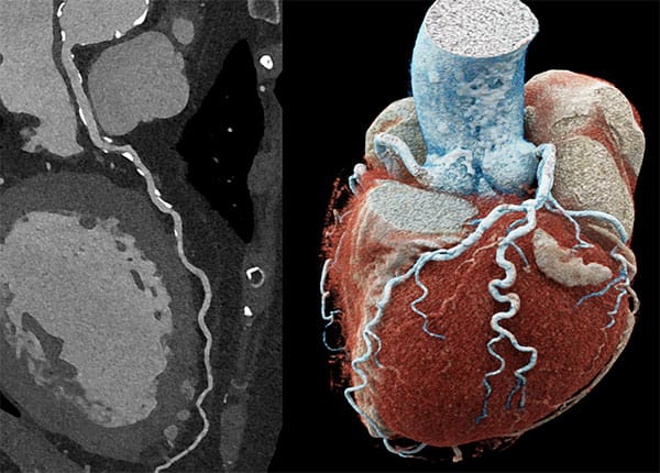
In a recent meta-analysis, stenosis was downclassified in severity in 34.2% of vessels when scanning with PCCT compared with conventional CT (95% CI, 27.3-41.9; P < .0001). The study authors attributed this to a reduction in blooming and better resolution. The study also reported fewer downstream invasive CAG referrals for PCCT scans than for conventional CT scans (pooled odds ratio [OR], 0.71; 95% CI, 0.63-0.79; P < .0001) and significantly higher rates of revascularization among those undergoing invasive CAG (OR, 1.43; 95% CI, 1.14-1.79; P = .0017).
Similarly, a single-center retrospective study comparing nearly 4000 PCCT scans with CT scans found lesser use of CAG with PCCT than with CT (9.9% vs 13.1% of the time; P < .001). PCCT patients tended to be referred more appropriately because they were more likely to ultimately undergo revascularization than patients scanned with conventional CT.
PCCT: A Pricey Integration to Clinic
The FDA first cleared the use of PCCT in September 2021, a few months after the first human cardiac scan was met with a round of applause by researchers at Mayo Clinic in Rochester, Minnesota. Shiyovich estimated that about 100 PCCT scanners are now operational across the US and about 200 are now operational around the world. He expects PCCT to become the gold standard within a decade or two.
In spring this year, the FDA cleared the first portable PCCT designed for musculoskeletal imaging in the community by a New Zealand-based medical device company. The FDA pointed to lower radiation, ultrahigh spatial resolution, sharp bone detail, and lower electronic noise offered by the scanner.
The $5 million sticker cost is still prohibitive for many sites. In addition, there is an upfront cost for the technological infrastructure needed to support higher-resolution image storage and the increased bandwidth it takes to transfer these images.
The PCCT Bottom Line
There are some imaging issues that PCCT does not solve, Shiyovich pointed out. PCCT can be performed with potentially lower radiation (for 0.4-mm scans), but the ultrahigh-resolution images (0.2-mm scans) require comparable radiation. He noted that PCCT does not eliminate the need for contrast, which is an issue for patients with allergies or kidney disease. “Breath holding and heart rate control still need to be considered,” he added.
For the clinician, there’s no added reimbursement and it’s a bit more complicated for the reader, Villines told Medscape Medical News. But aside from affordability, “there’s no downside. It’s very state of the art…. We are excited about it.”
Villines reported leading clinical trials for Abcentra, Cleerly, HeartFlow, and Milestone Pharmaceuticals. Shiyovich reported being a paid consultant for Artrya Ltd and receiving honoraria from Pfizer.