

 Admin_Adham
More than 8500 people are expected to die from melanoma in the US in 2026. Early detection dramatically improves survival, yet the tools for identifying suspicious lesions remain, well, cumbersome.
Dermoscopy requires specialized training and remains dependent on a trained operator. Reflectance confocal microscopy offers cellular-level detail but demands expensive equipment, often exceeding $50,000, and is largely confined to academic medical centers. And biopsy, the gold standard, is invasive, requires pathology review, and is often performed on lesions that turn out to be, fortunately, benign.
Now, researchers at Wake Forest University School of Medicine in Winston-Salem, North Carolina, and collaborating institutions have developed a flexible, battery-free patch that measures the bioimpedance of skin lesions wirelessly, potentially putting preliminary melanoma screening in the hands of primary care providers and patients themselves.
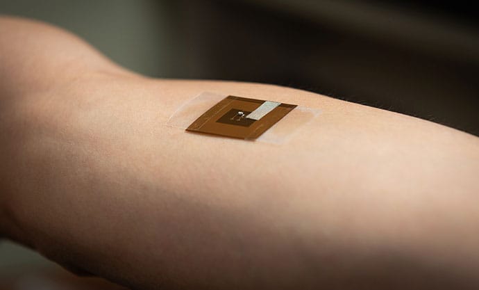
The proof-of-concept study, published in npj Biomedical Innovations, demonstrates that the wearable device can detect electrical differences between pigmented lesions and healthy skin, though substantial validation remains before it could reach clinical use.
“Our motivation was to make a device that is accessible to people that they can use at home for screening melanoma and skin cancer for early identification and follow-up medical attention,” said Mohammad J. Moghimi, PhD, assistant professor of biomedical engineering at Wake Forest University School of Medicine and the study’s senior author.

For Moghimi, whose training is in electrical engineering, the project represents a convergence of disciplines. “Wireless communication, things that you learn in college — all the pieces come together,” he said. “My background in electrical engineering and my discussions with medical doctors significantly helped develop this device.”
The Access Gap
The principle behind bioimpedance sensing isn’t new. Cancer cells exhibit different electrical properties than healthy tissue. Higher perfusion, altered ion concentrations, different pH, and elevated temperature all contribute to higher conductance and lower impedance in malignant tissue.
Electrical impedance spectroscopy (EIS) devices already exist and have demonstrated clinical value: Studies have shown that adding EIS to dermatologic evaluation increased correct biopsy decisions from 59.9% to 71.0%, with nearly 40% relative improvement in diagnostically challenging cases.
But existing EIS devices come with significant barriers. SciBase’s Nevisense, one of the most established FDA-cleared impedance-based systems, is a benchtop device costing thousands of dollars. It uses penetrating micro-electrodes that puncture the skin, requires specialist operation, and is designed for clinical settings rather than home use.
“Most devices that measure skin bioimpedance with high resolution are very expensive,” Moghimi explained. “They are minimally invasive and require specialists to run the equipment. Our idea was to address these issues.”
The patch his team developed takes a fundamentally different approach: a passive, wireless design that eliminates batteries, chips, and invasive electrodes. It’s a rethinking of how impedance sensing could work by miniaturizing existing technology and reimagining the architecture from the ground up.
Reading Skin, Wirelessly
The system consists of two components: a flexible patch that goes onto the skin and a separate reader module. The patch, which is about an inch square and thinner than a credit card, contains a copper inductor coil and capacitor printed on a polyimide substrate. Surface electrodes contact the skin without penetrating it.
The reader module, held just a few millimeters from the skin, powers the patch wirelessly — similar to tapping a phone to make a payment. Once powered, the patch sends a tiny electrical current through the skin and measures how the tissue responds. Different tissues resist electrical flow differently, and that difference shows up as a shift in the signal the reader picks up.
The simplicity is by design, Moghimi said. “You tap your phone or the external module on the patch and read the data. So you don’t need a battery. It’s much easier, the device is thinner, and the cost is significantly reduced when it’s passive.”
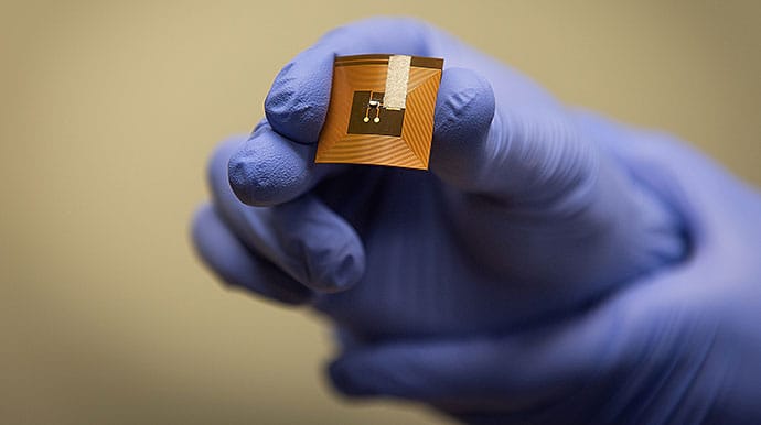
The battery-free design eliminates several practical barriers to home use. Batteries add bulk, require charging, degrade over time, and increase cost. By drawing power wirelessly only when measurements are taken, the patch can be thin, flexible, and potentially disposable.
Crucially, the system measures impedance of the lesion relative to healthy skin 2-3 cm away on the same patient — not absolute values. This internal reference approach eliminates confounding variables like ambient humidity, skin moisture, and individual variation in baseline electrical properties.
“We measure impedance of each person at the lesion compared with their own healthy tissue,” Moghimi explained. “It’s not sensitive to skin tone. You only look at the difference rather than absolute values.”
From Grocery Store to Human Trials
Moghimi’s team first validated the physics with dummy electrical loads in the lab. Then came an unlikely next step: A graduate student walked into a grocery store, bought some chicken, and brought it back to test.
“I think that was the moment we were very confident,” Moghimi recalled. “We could see the shift in the resonance frequency, and we were able to compare it with measurements from benchtop equipment. They followed the same trend.”
From there, the team progressed to human individuals. They tested the patch on 10 volunteers, measuring the impedance of pigmented lesions (moles) compared with adjacent healthy skin. The results were consistent across all individuals.
The electrical differences showed up in two key measurements. The first measurement was capacitance, which is a measure of how much electrical charge tissues can store, related to cell membrane properties.
Moles showed higher mean capacitance (7.3 picofarads) than healthy skin (3.8 picofarads), suggesting differences in cellular structure and membrane composition.
The second measurement was resistance, which is how much the tissue opposes electrical current flow, influenced by ion concentration and fluid content. Moles showed lower mean resistance (4.5 kilohms vs 8.3 kilohms), consistent with the higher conductivity expected in more metabolically active tissue.
The patch also detected differences in how much energy tissue absorbed vs reflected. Healthy skin absorbed more energy, while moles bounced more signal back. This gave the team yet another metric for distinguishing tissue types.
The results were consistent. Repeat measurements clustered tightly together, and when the team compared their patch to Nevisense — the FDA-cleared clinical device — the trends aligned, even though the raw numbers differed due to the devices’ different approaches to measuring skin.
The View From the Clinic
Steven Xu, MD, MSc, Medical Director of the Querrey Simpson Institute for Bioelectronics at Northwestern University in Chicago and CEO and co-founder of Sibel Health, offered perspective on where the technology fits in the diagnostic landscape (Xu was not involved in the study.)
“Using impedance is not new. It’s not new in skin. It’s not new in medicine,” Xu said. “What is new is [they] put [the patch] into this form factor that is different, which is more conducive to a wearable, that could potentially be continuous. There’s an evolution of the solution here, where the form factor creates new clinical workflow benefits.”
Xu noted several potential advantages: the possibility of home monitoring without clinic visits, longitudinal tracking of suspicious lesions over time, and reduced burden on specialist referral systems. For patients with dysplastic nevus syndrome, who may have hundreds of moles requiring surveillance, a low-cost, take-home option could meaningfully change their care experience.
But he also outlined the validation challenges ahead. “You don’t want to miss a melanoma on your skin because that’s really, really bad, so you need to be very sensitive,” Xu explained. “But if you tell me everything’s a skin cancer, that’s not helpful either — you have pretty low specificity” and unnecessary biopsies.
Existing technologies have grappled with this balance. Nevisense achieved approximately 97% sensitivity in clinical trials but with lower specificity. MelaFind, another FDA-cleared device that used multispectral imaging, faced criticism for false-positive rates and is no longer on the market.
For the patch to demonstrate clinical value, it would need to approach similar sensitivity benchmarks while ideally improving on specificity. The wearable format could potentially help here — multiple readings over time might reveal patterns that single measurements miss.
Xu also raised questions about clinical workflow. Melanoma screening is typically a point-in-time decision — biopsy or not — rather than a condition requiring continuous monitoring like blood glucose or heart rhythm. Whether a wearable adds value for spot-check decisions remains to be seen.
“I think there are ways this could help — tracking lesions in sensitive places, increasing access,” Xu said. “These are things where the form factor could really make a difference.”
More broadly, Xu sees the future of wearables in dermatology less in diagnosis than in prevention and monitoring, like tracking scratching behavior in patients with eczema, sensing ultraviolet (UV) exposure before sunburns happen, and measuring skin barrier changes with the seasons.
Equal Performance Across Skin Tones
The paper highlights that impedance sensing works equally well across all skin tones, unlike artificial intelligence [AI] imaging systems trained predominantly on lighter skin that have shown documented bias. Because the patch measures electrical properties rather than visual features, melanin content doesn’t affect readings.
Xu offered context: While equity in diagnostic technology matters, the epidemiology of melanoma skews heavily toward lighter-skinned individuals with high UV exposure. When melanoma does occur in darker-skinned patients, it often appears on acral sites — palms, soles, and nail beds — that some devices specifically exclude.
“Yes, AI equity, [those types] of things are very important,” Xu said. “But we also need to understand the actual disease burden and where these tools will be used most.”
Still, validation across diverse skin types will be essential, and a technology that works regardless of pigmentation removes one variable from an already complex diagnostic picture.
From Bench to Bedside
Moghimi’s team is moving toward larger clinical trials at Wake Forest’s Comprehensive Cancer Center, this time testing the patch on actual melanoma lesions rather than benign moles. They’re also exploring applications beyond melanoma: nonmelanoma skin cancers (basal cell and squamous cell carcinoma), wound healing monitoring, and potentially mental health applications through skin conductance measurement.
The regulatory pathway appears relatively straightforward given the device’s noninvasive nature and low electrical output, but Moghimi estimates clinical availability is “probably years” away, pending robust safety and efficacy data.
The team envisions two parallel use cases: a consumer device for home screening that could alert users to seek medical attention and a clinical tool for primary care providers to help triage referrals to dermatology.
“Primary care doctors, when they see a mole, sometimes they don’t know whether it’s high risk or not from appearance,” Moghimi said. “This could help them identify which lesions are more likely to need specialist evaluation.”
“Something that’s accessible to people can potentially save lives, reduce the cost of treatment, and reduce the burden of disease,” Moghimi said. “That’s the mission of my research.”
Moghimi reported having an international patent cooperation treaty application on wireless bioimpedance measurement with wearable patches.
