Admin_Adham
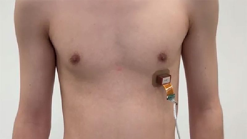
Pacemakers have become smaller and more sophisticated over the decades, but they still share one defining characteristic: They must enter the body to operate. Researchers at the Massachusetts Institute of Technology (MIT), Cambridge, Massachusetts, challenge that technology with a wearable, noninvasive pacemaker that stimulates the heart using ultrasound from outside the body.
The study, published in Nature Biomedical Engineering, demonstrated how the tiny, postage-stamp-sized device could synchronize contractions in engineered human heart cells, restore normal heart rhythms in animal models, and maintain a favorable safety profile during 8 months of follow-up.

Seeing the technology work in a living system was a milestone years in the making. “It was an exciting and rewarding moment,” said Chen Gong, PhD candidate and first author of the study at MIT. “The project required years of work across multiple disciplines, and seeing the heart respond reliably to wearable ultrasound stimulation was the first confirmation that this concept could work in a living system.”
A Different Way to Reach the Heart
Pacemaker technology is one of medicine’s great success stories. But despite decades of innovation, it remains inherently invasive. Even leadless pacemakers, which eliminate many of the complications associated with traditional leads and surgical pockets, must still be implanted inside the body through a minimally invasive procedure. The desire to reduce the burden of implantation has fueled a wave of innovation across cardiac rhythm management. Conduction system pacing has gained attention for delivering electrical stimulation that more closely mimics the heart’s natural activation patterns.
Scientists are also exploring entirely new technologies. A 2025 study published in Natureby a team at Northwestern University introduced a temporary pacemaker, small enough to be injected into the body and designed to dissolve once it’s no longer needed. Other groups are investigating biological pacemakers that use living cells to restore rhythm without the need for permanently implanted devices.
The MIT team’s noninvasive pacemaker occupies a unique position because it seeks to eliminate implantation altogether by combining wearable bioelectronics with genetic engineering. The answer, they believe, lies in ultrasound and sonogenetics. “Our sonogenetic approach uses wearable ultrasound to stimulate genetically sensitized cardiac cells from outside the body, potentially offering a less invasive alternative in the future,” Gong said.
Teaching Cells to Hear Sound
The idea of using ultrasound to influence the heart is not new. Researchers have long known that acoustic waves can affect cardiac tissue, and ultrasonics have been explored as a potential tool for noninvasive pacing. The problem was reliability. Previous studies generally produced effects that were too weak or inconsistent to control the heartbeat in a predictable way.
Sonogenetics, an emerging field, uses genetic engineering to make cells more responsive to sound. The team introduced the mechanosensitive ion channel MscL-G22S into human cardiomyocytes derived from embryonic stem cells. When exposed to ultrasound pulses, the cells displayed synchronized calcium signaling and contractions. Unmodified cells showed far less responsiveness.
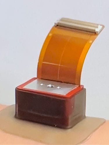
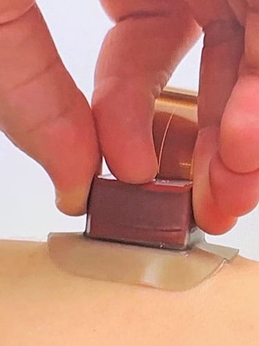
The team then incorporated the technology into a wearable noninvasive ultrasound pacemaker, which consists of a hydrogel-based patch embedded with miniature ultrasound transducers that stick to the chest.

“In the rat experiments, we saw animals with slow heart rates brought up to normal, and animals with irregular heartbeats steadied so that the heart kept in sync with ultrasound’s ‘ticks,’” said Xuanhe Zhao, PhD, professor of mechanical engineering and of civil and environmental engineering at MIT. The team also reported chamber-specific pacing with submillimeter spatial precision and frequency control of up to 9 Hz.
Just as important, no significant adverse effects were observed during 8 months of follow-up. The team also demonstrated successful stimulation in ex vivo porcine hearts. This provided additional evidence that the noninvasive pacemaker remains feasible as heart size increases.
Achieving those results proved considerably more difficult. “One of the biggest challenges was achieving reliable heart rhythm control while keeping the system completely noninvasive,” Gong said. The team had to optimize gene delivery, ultrasound parameters, and device design simultaneously while accounting for the heart’s constant motion to ensure precise and repeatable stimulation.
The Challenges Ahead
The technology’s biggest challenge may also be its defining feature. The current system depends on the genetic modification of cardiac cells. Before ultrasound pacing can occur, cardiomyocytes must express the engineered ion channel that allows them to respond to acoustic stimulation.
“A key limitation is that the approach currently requires gene delivery to the heart,” Gong acknowledged. “Future work will focus on improving gene delivery methods, evaluating long-term safety, testing in larger animal models, and developing more advanced wearable ultrasound systems for potential clinical translation.”

Both the gene therapy and the ultrasound delivery system face significant hurdles, said Kenneth Ellenbogen, MD, cardiac electrophysiologist and Kimmerling Professor of Medicine in the Division of Cardiology at VCU Health Pauley Heart Center in Richmond, Virginia. He was not involved in the research.
“It is a very interesting approach, but it is a very big step forward that will require major further breakthroughs in ultrasound transmission,” he said. Human anatomy could complicate matters because the ultrasound transducer may be physically distant from the targeted cells. He also noted that “gene transfer of the transfected cells presents a formidable challenge,” particularly because the durability of the response remains unknown.
Ellenbogen added that the system’s long-term performance remains largely unexplored and requires significant advances to make the pacemaker a reality. “It appears that this approach could be used for most pacemaker patients, but its suitability for pacemaker-dependent patients — that is, patients whose hearts cannot maintain a normal rhythm without a pacemaker — would need to be studied in more depth,” he said.
A Glimpse of the Future
The wearable ultrasound pacemaker is still in the preclinical stage and significant work remains. “Before human trials, we need additional studies on long-term safety, gene delivery, and validation in larger animal models. Human applications are still several years away,” Gong said.
Even so, the concept raises intriguing possibilities for the future of cardiac rhythm management. According to Ellenbogen, a noninvasive ultrasound pacemaker could reduce risks associated with permanently implanted hardware, such as infection, bleeding, tissue injury, and scarring.
Perhaps the most remarkable aspect of the MIT study is how it reimagines a technology that has remained fundamentally unchanged for decades. It offers a glimpse that the pacemakers of the future may still regulate the heartbeat in much the same way as today’s devices, but they won’t need to be inside the body to do it.
Ellenbogen reported having no relevant conflicts. Disclosure information for study authors is available in the original study publication.