Admin_Adham
Macrophages can strip tiny parcels of cytoplasm from living cells, shelter the material from degradation, and use it to activate CD8 T cells — the cytotoxic arm of adaptive immunity.
A team at the University of California, San Francisco (UCSF), used high-resolution microscopy to capture macrophages pinching off submicrometer vesicles from healthy neighbors without killing them. This is a version of trogocytosis, a process in which a cell takes a bite of another cell’s membrane and contents and leaves the rest intact.
The study, published in April in Nature, traced the nibbled cargo into a compartment that escapes the lysosome, where cells normally digest what they swallow. Sheltered from destruction, the sampled material was loaded onto major histocompatibility complex (MHC) class I — the molecule that flags a cell’s contents for inspection by CD8 “killer” T cells. Displaying outside material this way is a feat usually reserved for dendritic cells, the immune system’s professional teachers. And the effect was specific: the nibbled material switched on CD8 T cells while leaving the CD4 “helper” T cells untouched.

“My reaction was that this cannot be right,” lead author Amy Fan, PhD, said, describing the first time her flow cytometry data showed macrophages activating CD8 T cells from live-cell material. She had colleagues repeat the experiments before she accepted the result.
“This is a way for the immune system to accumulate information about our healthy self,” said senior author Matthew Krummel, PhD, professor of pathology at UCSF. While the idea that immune cells sample living cells had been documented, no one had been able to watch the act itself unfold at this scale, said Krummel.
Julie M. Blander, PhD, Gladys and Roland Harriman Professor of Immunology in Medicine at Weill Cornell Medicine in New York City, who was not involved with the study, called the results a genuine advance. “One of the commandments of basic immunology is that macrophages don’t cross-present to CD8 T cells,” she said. “And here’s this paper showing macrophages can do it by nibbling not from dead cells, but from living cells. It does open new conceptual frontiers.”
An Easy Process to Miss
Trogocytosis is an ancient process, first glimpsed in the 1970s when a microbiologist watched the brain-eating amoeba Naegleria fowleri nibble mouse cells piecemeal rather than swallowing them. Researchers gave the process its name in 2003, and in the decades since, it has turned up across the immune system — in T cells, B cells, natural killer cells, neutrophils, dendritic cells, and macrophages — though its purpose has remained, in many contexts, unclear.
The UCSF team engineered mouse tissues — lung, skin, and intestinal lining — to glow green with a fluorescent protein called ZsGreen. When they looked at the macrophages sitting in those tissues, the macrophages were glowing too, loaded with tiny green specks.
The observed signal was specific to certain tissues. For example, macrophages in the lung carried lung protein, but macrophages in the spleen did not. And when the team transplanted normal bone marrow into the mice exhibiting the green fluorescence, the transplanted immune cells lit up with the host’s protein, proving they were picking it up from their neighbors.
We stared at each other and were like, this looks real.
To watch the process more closely, the team cultured macrophages alongside green-labeled melanoma cells or fibroblasts. The macrophages took up green signal at intensities hundreds of times dimmer than the intact donor cells, as though they had sampled a sliver rather than swallowed a whole cell. Whatever the macrophages were acquiring, they had to touch the cell to get it.
The researchers wondered: Are these dying cells? To test that, team pharmacologically blocked cell death (apoptosis), yet the macrophages continued to nibble the cells.
How They Witnessed the Act
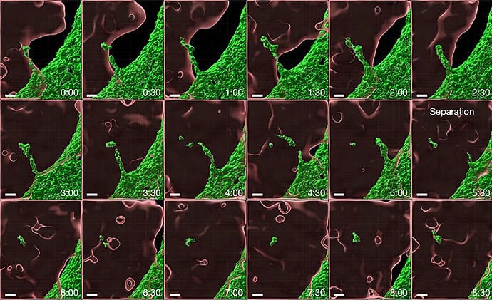
To watch the event in real-time at miniscule resolution, the team used two separate high-resolution systems: lattice light-sheet microscopy, which captures full cell volumes over time with minimal photodamage, and a Nikon spatial array confocal detector. In the study’s videos, a protrusion of the target cell stretched into the macrophageover the course of < 10 minutes. The decisive moment — the actual abscission of the vesicle — happened within a single 30-second frame. The separated vesicles were tiny, ranging from 0.02 to 0.05 µm3, on the order of a thousandth of a cell’s volume.
Krummel compared the challenge of imaging the event to wildlife cinematography: “It’s like a National Geographic photo shoot that requires you to be under the blinds for hours,” he said. The team spent days in a dark room, capturing video after video, hoping to catch a complete event, according to Fan.
When the team sorted macrophages that had taken up fluorescent material and imaged them, multiple people in the lab examined the data independently. “We stared at each other and were like, this looks real,” Fan said. “This doesn’t just look like a blip that came up and went away.”
To trace what happened to the cargo after uptake, the team developed an organelle profiling method that could identify whether sampled material was sitting in early endosomes, late endosomes, lysosomes, or recycling compartments. The method revealed a stark difference. About 90% of vesicles from ordinary phagocytosis of dead cells carried a conventional endocytic marker. But a substantial share of live-sampled vesicles carried none, so the nibbled material was going somewhere else.

They then found a gene involved in directing where the nibbled material goes, called Snx27. When they deleted it, the macrophage’s nibbled cargo got dumped into the cell’s digestive machinery instead of being held aside, and CD8 T cells stopped responding. But the macrophage itself was fine: hand it a premade antigen fragment and it could still do the job.
And the route was selective. Macrophages that sampled live cells activated CD8 T cells. Macrophages that ate dead cells did not.
“They are basically factories for digestion and clearance,” Blander said of macrophages in general. Yet the live-sampled material avoided that fate. “They internalize things, and immediately they deliver things to lysosomes for degradation, so they’re no longer going to preserve the epitopes for class one presentation.” The nibbled material, by contrast, was being held in reserve. “In terms of macrophage biology and the spectrum of cells that can cross-present, it’s not just dendritic cells doing this all the time.”
Is It New, or Newly Visible?

Daniel Fletcher, PhD, professor of bioengineering at the University of California, Berkeley, offered independent confirmation that the process is not exotic. His lab published a study in Nature Cell Biology showing that a target cell’s cortical tension (the mechanical stiffness of its outer surface) governs whether a macrophage nibbles or swallows.
“If you really look for trogocytosis, it’s going on all the time,” he said. “It’s happening so much. I think that was surprising.”
This nibbling in trogocytosis sets what he called the ‘tone’ of the immune system, a running census of healthy self.
He called the trafficking and CD8 results the most significant part of the paper. “Their identification of key factors in avoiding trafficking to the lysosome is really exciting, and the fact that it is used to signal to CD8 T cells — that’s super cool,” he said.

Krummel ventured a more speculative reading of his own data: that this nibbling in trogocytosis sets what he called the “tone” of the immune system, a running census of healthy self that tells T cells what to ignore. Immune tolerance (the process by which the body learns not to attack its own tissues) has been shown to be maintained largely through CD4 regulatory T cells and enforced by tissue-resident immune populations that live in the organs they protect.
Blander said Krummel’s framing is a viable interpretation. “People think of cDC1s (a subset of dendritic cells), but if you look intratumorally, there are many more macrophages, and they’re all thought to be suppressive,” she said. “Are they doing something to tolerize the CD8 T cells even more? You don’t really need ongoing apoptosis as a source of self-antigen continuously…you could also have nibbling from live healthy cells, just for our bodies to say, ‘this is us.’”

Peter van Endert, MD, PhD, an immunologist at Université Paris Cité and INSERM in Paris, France, who was not involved with the study, said, “The technology here is more novel than the biology…Live trogocytosis by myeloid cells has been seen clearly before.”
He noted Anne Hosmalin’s group published a study in 2010 showing that dendritic cells take up antigen from healthy live cells in mice and cross-present it, with protective immune responses in vivo. What this new study adds, in van Endert’s reading, is resolution, and the extension from dendritic cells to macrophages.
He also questioned the leap from the experimental system to the immune tolerance claims the paper makes. The macrophages in this study were bone-marrow-derived. “They’re closest to inflammatory macrophages — what you find in the peritoneal space when you irritate it, such as injecting stuff into a mouse,” van Endert said. “You wouldn’t think that they’re very likely to contribute to immune tolerance.”
He noted the paper’s makes tolerance claims about CD8, the killing arm, not the regulatory one. “It’s a step too far,” he said. “I don’t know if I accept that this would contribute to immune tolerance.”
The Open Questions Worth Chasing
Van Endert flagged one of the paper’s largest gaps: the identity of the compartment where the sampled antigen is kept away from the lysosome. “They don’t really explain how,” he said. He raised a mechanistic objection too. “The receptors that are involved, CD11b and CD93, are the receptors that do classical phagocytosis,” he said. “So how could the cell distinguish whether what’s internalized comes from a live cell or from dead-cell debris? That I don’t understand mechanistically.”
Fletcher’s study investigated what determines whether a cell distinguishes between dead-cell debris vs live cell for cell-nibbling from a biophysical angle — cortical tension — so the receptor question is still open.
Blander was struck that the team had not tested for Rab11a, a molecular switch involved in vesicle trafficking that, in her own work, stocks a recycling compartment with large reserves of MHC class I, the molecule that displays intracellular antigens to CD8 T cells. If sampled antigen lands there, it arrives exactly where the loading machinery waits. “My bet is that it’s Rab11a positive,” she said.
However, Fan is now hunting for a positive marker of that compartment using low-input mass spectrometry.
Because macrophages don’t travel to lymph nodes, where naive T cells are trained, Blander suggested the sampled material either reaches CD8 T cells already in the tissue or gets handed to a dendritic cell that can migrate and carry the lesson forward. She also wondered whether the pathway survives infection at all. “When a macrophage is activated, Toll-like receptors speed up phagocytosis and maturation into lysosomes,” she said. “So retaining things in a nondegradative compartment may not be operational when you have an infection. I wish they had shown that.”
“If live-sampling is a consistent source of antigen in vivo,” Fan said, “it reframes how macrophages may be tuning T cell responses, what we should be measuring to understand it, and how we might target those interactions.”
“[Krummel’s team] is not looking at some anomalous condition or disease state,” Fletcher said. “He’s looking at regular background processes.”
Fletcher, van Endert, and Blander reported having no relevant disclosures. Disclosure information for study authors is available in the original study publication.