
 Admin_Adham
 Jump to Key Points
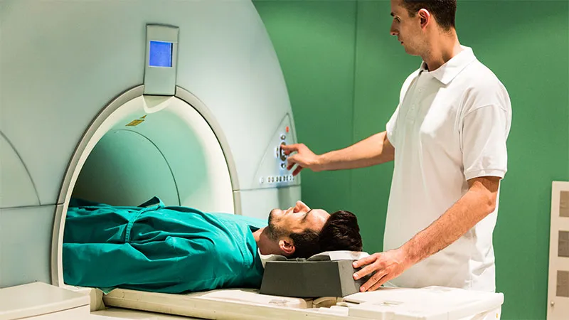
LAS VEGAS — The task of distinguishing axial spondyloarthritis (axSpA) from the myriad underlying causes of back pain has become largely dependent on obtaining multiple targeted MRI sequences, meaning that rheumatologists ordering these images should understand the principles of MRI, not just leave it to the radiologist.

“I can tell you that you are working with both hands tied behind your back if you are not familiar with how to employ MRI in the classification of axSpA,” said Walter P. Maksymowych, MD, a rheumatologist and professor of medicine at the University of Alberta in Edmonton, Alberta, Canada.
The importance of MRI is reflected in the 2025 revised classification criteria jointly issued by the Assessments in Spondyloarthritis International Society (ASAS) and the Spondyloarthritis Research and Treatment Network (SPARTAN).
Relative to a prior set of classification criteria issued in 2009, the sequence of images increased specificity from approximately 80% to more than 90%.
- axSpA dx/classification now heavily MRI-dependent; specificity >90% with revised criteria.
- SIJ MRI requires multiple targeted sequences; lumbar MRI alone is insufficient.
- BME alone is nonspecific; postpartum women/athletes may show false-positive load-zone lesions.
- SASPIC/CLASSIC: MRI added major diagnostic value vs clinical/lab/radiographic data alone.
- Imaging confirmation advised before biologic/targeted synthetic DMARD initiation.
In a presentation delivered at Perspectives in Rheumatic Diseases (PRD) 2026, Maksymowych called specificity rates of less than 90% “simply not good enough” given the high prevalence of unexplained back pain and the risk of prescribing therapies to patients who will not benefit.
Very Specific MRI Sequences Needed for axSpA
The point of encouraging clinicians to develop a more comprehensive understanding of MRI is that “you cannot just order a lumbar-spine MRI and expect to get the information you need,” Maksymowych explained. He provided several examples of the risks for false positives or false negatives if the most up-to-date MRI sequences, such as those involving short tau inversion recovery for fluid-rich tissues, are not ordered.
In a case example, he cited a report on a 29-year-old woman with a 1-year history of back pain but a normal level of C-reactive protein (CRP). Based on a T1-weighted scan showing fat metaplasia in the anterior sacrum and evidence of bone marrow edema (BME), the report called findings “suspicious for sacroiliitis.”
Yet with the greater precision of imaging conducted on both the semicoronal and semiaxial plane, BME was found confined to the load zone, which Maksymowych described as a “red flag” for false-positive axSpA.
In postpartum women, as in this case, as well as in some athletes, focal BME in the load zone does not constitute axSpA and would not be helped by an anti-inflammatory biologic. Rather, he said physical modalities were the appropriate therapy in this case.
“I have looked at the very first scan of patients who have cycled through five biologics only to find that axSpA was the wrong diagnosis,” Maksymowych said.
Multiple MRI Images Needed for axSpA
Yet no single MRI image will rule in or out axSpA. Rather, Maksymowych said clinicians need to have enough understanding of MRI to weigh which sequences to order for disease activity in the anatomically complex sacroiliac joint (SIJ). The best sequences to detect inflammatory lesions, fat metaplasia, BME, bone erosion, and other signs of axSpA differ.
“MRI indicative of axSpA is a synthesis of information from all MRI sequences,” Maksymowych said. Based on a 2025 review from his own institution, he said a standardized methodological approach is appropriate to gather this information. It must be evaluated in the context of clinical symptoms and other diagnostic studies, such as laboratory evidence of elevated systemic inflammation.
But Maksymowych believes “MRI has a profound impact on diagnostic accuracy” and should now be included in the workup of all patients suspected of axSpA.
The supportive evidence is drawn from the two-center SASPIC study Maksymowych conducted in Canada and the subsequent CLASSIC study, which ultimately led to the new 2025 ASAS-SPARTAN classification criteria.
In the SASPIC study, 373 patients presenting with back pain and one of three inflammatory diseases — psoriasis, inflammatory bowel disease, and acute anterior uveitis — were screened in a structured diagnostic evaluation for axSpA. The value of diagnostic studies was evaluated separately in B27-positive and B27-negative patients for each of the three inflammatory disease groups.
The study looked at the added value of MRI when compared to either clinical signs alone or to clinical signs plus positive laboratory and radiographic evidence. The gold standard was a diagnosis made on the basis of clinical signs, laboratory evidence, radiological evidence, and MRI. Diagnostic accuracy was substantially enhanced with MRI compared to any other set of diagnostic evidence alone or in combination.
For example, the diagnosis of axSpA was reached in only about 15% of patients with psoriasis who were B27 negative with the gold standard of diagnostic studies when MRI images were read centrally. In comparison, 70% would have qualified for an axSpA diagnosis on clinical signs and more than 50% on the other criteria without MRI.
Notably, the 15% rate of a confirmed axSpA diagnosis with a gold standard workup was about half of the approximately 30% rate produced when the gold standard workup involved locally read rather than centrally read MRI.
Other notable findings from this study included the fact that only about 50% of those who received a confirmed diagnosis on the basis of the gold standard workup were B27 positive. No clinical features were found useful for distinguishing those with confirmed axSpA from others in the study. Centrally read MRI scans reduced the rate of confirmed axSpA regardless of B27 status in the psoriasis and IBD groups. They were similarly accurate in the uveitis group.
CLASSIC Study Also Supports MRI
In the CLASSIC study, there was a similar evaluation of axSpA diagnosis when adding clinical assessment, laboratory measures, pelvic radiographs, and local MRI assessments. When the 370 patients with confirmed axSpA were compared to the 645 patients for whom axSpA was ruled out, MRI evidence of axSpA was the most powerful diagnostic variable.
For example, 39% of those with confirmed axSpA had elevated CRP, defined as > 5 mg/L, relative to 23.3% of those without, but MRI indicative of axSpA increased the rate of confirmed axSpA regardless if the scans were read locally (81.9% vs 12.1%) or centrally (61.6% vs 1.7%) even if P values for these differences were not reported.
The most comprehensive global assessment evaluated in CLASSIC included clinical features, laboratory measures, radiographs, and MRI but did not reach a goal specificity of 90%. The specificities were 84.3% and 77.1% for MRI read centrally and locally, respectively. However, the 2025 ASAS-SPARTAN revised classification based on these data did meet this goal.
This approach employs a weighted point system with a cutoff for axSpA classification of 11 points or greater. Most clinical criteria, such as elevated CRP or peripheral arthritis, receive a single point, meaning that the 11-point threshold is difficult to reach without either MRI evidence (8 points) or radiographic evidence (7 points). In this system, the only other characteristics awarded more than 1 point are a positive HLA-B27 (4 points) and inflammatory back pain (3 points).
In the event of negative HLA-B27 status and negative imaging (both MRI and radiographic), the system does not permit classification. In those with positive HLA-B27 status but negative imaging, the 11-point threshold is met only when all other clinical criteria are met, but this requires multiple sites of inflammatory disease involvement.
For those who achieve or exceed 11 points, specificities for axSpA classification reached 89.2%-90.4% when MRI was interpreted by a local reader but were higher at close to 99% when MRI was read centrally and two of three central readers agreed on the MRI component. The sensitivities were nearly 80% with MRIs interpreted by a local reader but around 60% when interpreted by central readers, Maksymowych reported.
Biologics Should Not Be Started Prior to MRI
Based on these data, “I never initiate a biologic or targeted synthetic DMARD without imaging confirmation,” Maksymowych said. The risk for a false-positive diagnosis is that “you put a patient on a biologic, perhaps in some cases for decades for the wrong reason.”
For those who are not confident about their expertise in MRI, Maksymowych recommended pursuing a course on this topic or otherwise improving skills.
“I strongly recommended that you do that. Your patients will love you for it,” he said.
Without a definitive test, the challenge of diagnosing axSpA with a high degree of specificity and sensitivity has long involved a synthesis of clinical, laboratory, and imaging findings. Of these elements, Torsten Diekhoff, MD, a senior consultant in the Department of Radiology at Charité – Universitätsmedizin Berlin in Berlin, agreed imaging in general is often essential for confirming the diagnosis.
However, in the context of a “well-intentioned push by rheumatologists” toward earlier diagnosis and treatment of axSpA, Diekhoff said the quality of the sequence of images cannot be compromised. A co-author of a 2025 Viewpoint in TheLancet Rheumatology on “the imaging crisis in axSpA,” Diekhoff, like Maksymowych, emphasized the role of “robust and reliable imaging criteria” to avoid overdiagnosis.
Despite the growing sophistication in which the SIJ can be evaluated with MRI, any finding alone, such as BME, is nonspecific, according to Diekhoff. Like Maksymowych, he also recommended a collaboration with rheumatologists to order the most helpful sequence of images.
“MRI is an essential and extremely powerful tool in the diagnosis of axSpA, but it needs to be used appropriately. It is important to interpret findings in the clinical context,” Diekhoff told Medscape Medical News.
“A crucial advantage of modern erosion-sensitive sequences is their ability to help us to question or reject a suspected diagnosis when other evidence is inconclusive. Therefore, a complete imaging protocol tailored to the SIJ is essential,” he said.
Speaking of the “fundamental conflict between diagnosing axSpA early and diagnosing it correctly,” Diekhoff urged clinicians to understand which images can inform other signs and clinical measures of axSpA to come as close to diagnostic certainty as possible prior to initiating chronic therapies.
Maksymowych reported having financial relationships with AbbVie, Alfasigma, Augurex, Bristol Myers Squibb, Janssen, Lakefront, Eli Lilly, Novartis, Pfizer, Quest Diagnostics, UCB Pharma, and Xbiotech. Diekhoff reported having financial relationships with AbbVie, Canon MS, Eli Lilly, Merck Sharpe & Dohme, Novartis, Pfizer, and UCB.
Ted Bosworth, a career medical writer based in New York City, has been covering advances in clinical medicine, including rheumatology, for several decades.